posted Thu, 12/22/2022 - 07:25
3913
+ 5
Pec tear MRI pics
ad
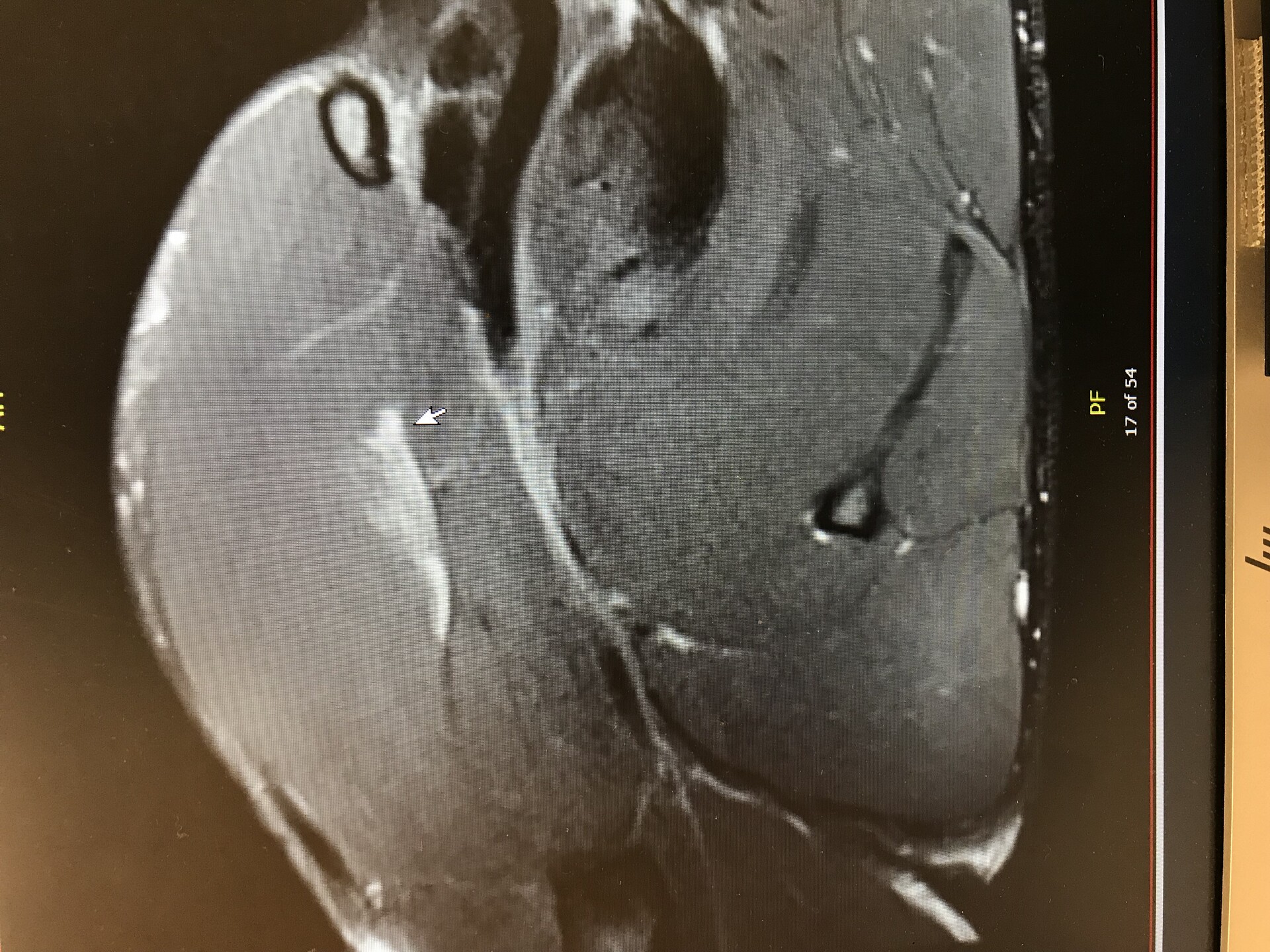
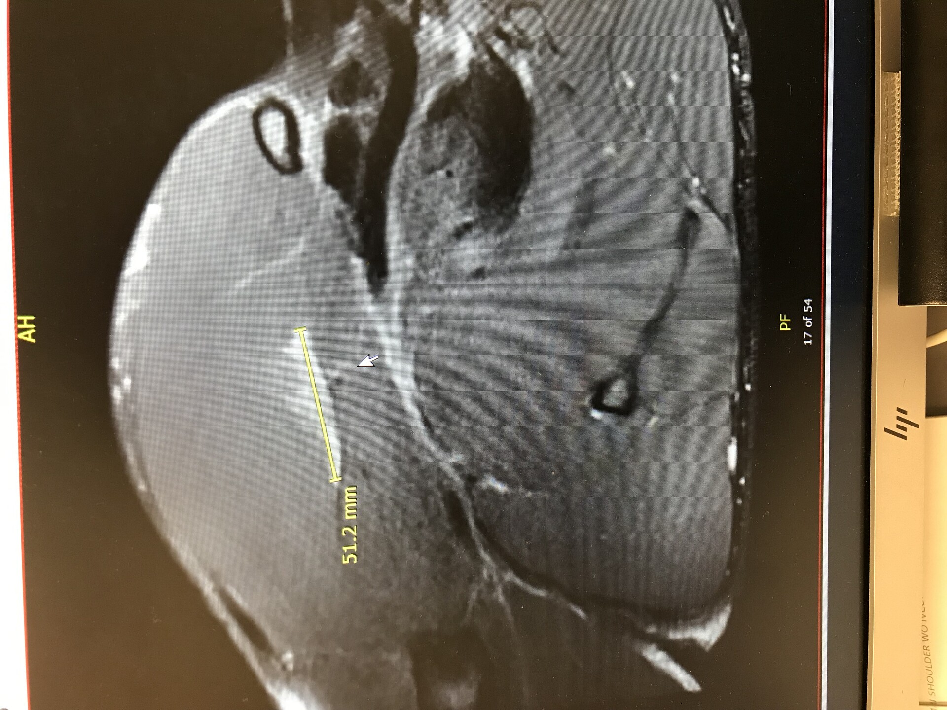
Pics are horizontal for some reason. Tap to enlarge and see it the right way.
The white line above the mouse cursor is the tear, and then it’s highlighted in yellow.
Partial pec tear from 7 weeks ago requested by @press1

GunShow22inI worked as an MRI Technologist for years and from only 2 images all I can say for sure is these are axial slices of the upper thorax and you appear to have a low amount of subcutaneous upper body fat;) Without being able to scroll through all the images its very difficult to orientate yourself.
I had a particial pec tear years ago and a small indentaton can be seen when I am sub 10% body fat and flexing my chest. I hope yours heals quickly and perfect unlike mine.
Get well soon. I guess you're on gh to heal. If not what's the protocol to heal it. I find local injection of bpc as close as possible to the injury help. It did for my lowe back. tb500 will help a lot too. It's effect is systemic so no need to be precise in the site of injection.
Bcp is very effective as a intranasel as well.
Had some bad elbow injuries, made self injection almost impossible.
Ai recover well bro very painfull ?'
I hope you get well soon
ShanntellsloverGet well soon
I can't figure out how I am looking at this here Lol Is that tear not on the front of the delt muscle? For reference what is the large circular hole?
Haha i cant read that at all lol im a little slow. I thought it was another pic of the dissolving grey stoppers on a sources bottle. But sounds like it hurts.
Your guess is as good as mine. Honestly I probably could’ve asked more questions. All I know is that as I was sitting in the office with the DR, she was highlighting that tear and saying this here is your pec major muscle and it’s torn right here. Don’t ask me the rest of what’s going on in that picture. According to her it’s torn in the upper portion belly of the muscle very close to where it connects near the armpit. But no it is not on the delt muscle whatsoever, but that’s what I thought as well when I first saw the picture lol. Im not a doctor I don’t even know what I’m looking at with these pictures other than the white line being a tear
I see exactly what your talking about. 50mm grade 2 tear right side mid upper pec. Only thing is it's not by the armpit like your Doc said.
My below comment is under normal hospital conditions.
So like I said below shame of your general Doc lol. The way it works is your MRI would of taken a bunch of visuals or images like you've taken and images are read by a Radiologist ( basically Doc whose specialty is Radiography). Radiologist knows anything before any one else would. That's what they do everyday all day...read those slides in the radiology dictation reading room.
Something to give you a laugh for the day Bud
https://www.youtube.com/shorts/B3Q88EGivvU
LMAO!!! Honestly mate it doesn't matter how many times I look at it I cannot make out what the different areas are in relation to your body. At one point I thought the hole may be where your heart is but that still doesn't make sense! Lol Maybe someone else who is more medical than us can work this out @HanginLow @Ghost
I have no idea what I am looking at here, this is way out my wheel house, hope for a quick recovery Klun
If you stair at it for 2 minutes then look away you see a picture of a lady form in mid air lol
hahaha It's like working out the bloody cripton factor isn't it!!! Who knew an exposed muscle could look so damn complicated to us muscleheads, if You look at a pectoral anatomy drawing it does seem to check out like Mak said.
If I am looking at this right it looks like the tear is in the clavicular head of pec. Your pec also partially covers what we would consider the front of the shoulder. You have the sternocostal head (front of your chest) and the clavicular head (shoulder region) which make up the entire pectoral muscle.
50mm tear on right side of mid upper chest from the look of the slides. Good it's not at insertion point. Looks between grade 2 and 3 tear.
Ahh Yes!!! I see exactly what you mean now Mak - I got an anatomy picture up breaking down the chest area's and see how it displays now. The MRI picture makes it all look strange eh lol
@Gh0st
Looks like hyper attenuation centrally suggesting inflammation/ partial tear at the origin (Sternum). This is non surgical.
Do you know what that black circle is mate?
Hard to tell exactly without being able to move through the MRI like you can on a program but based on location it’s likely the right atrium or ventricle of his heart filled with blood.
All very interesting stuff!
I prefer getting readings from Rad than looking myself when it comes to x rays of the chest but CT and MRI are not bad once you know what to look for.
Yes MRI are a little easier to read but he definitely should of gotten an explanation from a Radiologist, who actually would of made a complete diagnosis of the MRI slides. Lol shame on that Doc .
Good job we ain't surgeons eh!! lol
Interesting, Never had a pec tear, Is this something that will heal on its own or require surgery?
Due to mine being partial and in the belly of the pec major, should heal on its own. Luckily no tendons or points of insertions were involved. Should be able to recover without any visible defects. But right now still can’t even do a push-up without pain
Yea you were fortunate. Wish you a speedy recovery
Is that common to tear in the belly of the muscle. Seems like most would happen at insertion points.
From my understanding, very uncommon
Not that it is good, but probably better than tearing at an insertion I would think.
Oh Jesus yes!! Then he would have been in surgery like TRaww was.